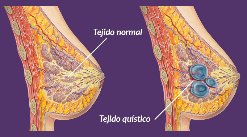
Fibrocystic breast disease
It is the most common benign breast disorder.
It constitutes 54% of mastopathies and 70% of benign lesions of the breast, they occur in the reproductive age of women, and therefore, the highest incidence is between 35 and 49 years.
Predisposing factors include: nulliparity (not having had children), late age at first delivery, high-fat diet, and obesity; considering breastfeeding as a protective factor. The origin of this disorder is functional, it responds to imbalances in female sex hormones favoring the appearance of breast cysts.
It consists of an increase in breast tissue.
Especially in the upper and outer areas of the breasts, towards the armpits, which makes them denser, forming cysts of variable size and number, it usually occurs in both breasts, although it can be of different intensity in one than in the other. In general, the patient reports discomfort in the affected breast, or both (mastodynia), or even intense pain (severe mastalgia), other times the patient does not report pain (asymptomatic) and is diagnosed after a breast ultrasound.
Regarding its cyclical characteristic, we must consider:
The diagnosis of this pathology is fundamentally clinical, that is, it is suspected due to the patient’s symptoms and the examination of the breast, it arises after a routine examination carried out by your doctor that upon examination highlights dense breasts, with irregularities that correspond to the presence of nodules or cysts that may or may not be accompanied by pain.
If fibrosis predominates, it is called fibrous mastopathy, or if both predominate, it is called fibrocystic mastopathy.
Auxiliary diagnostic methods may be necessary, such as mammography, breast ultrasound (ultrasound examination), aspiration of cysts, etc.
When a mass is clinically palpable and mammography detects a nodule with well-defined contours, ultrasound is what will allow its content to be distinguished between solid and liquid.
But if a mass is palpable and not seen radiologically, it must be removed by surgery and histopathologically studied.
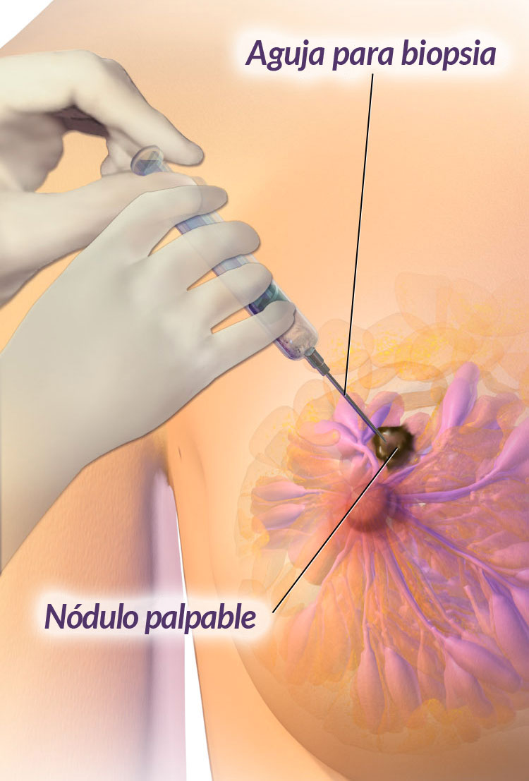
In the presence of a nodule with ultrasound signs of a cyst, the correct attitude will be the evacuating puncture to obtain material, which can be studied by cytology if it has bloody characteristics, or if it is cloudy or opalescent it should be sent for histopathological study. . If it is predominantly fibrous, the mammogram will show microcalcifications or calcifications, where it is already advisable to do a biopsy.
A palpable nodule in a woman with risk factors and/or ultrasound findings should be biopsied
Either fine needle or Tru-cut biopsy. If there are atypia, indicate excisional biopsy to rule out malignant processes present.
Taking into account the endocrine pathophysiology of fibrocystic mastopathy, represented by hormonal imbalance, medical treatment should not be single, but multiple. This constitutes the essence of combined treatment based on different hormones (progestogens) in conjunction with analgesics to relieve pain.
Individual treatment.
Treatment must be individualized and, on occasions, when the cysts are larger, punctures will have to be performed to aspirate the contents of the cysts and thus reduce them. Or if they are not palpable, they will require a fine needle aspiration or biopsy to rule out malignancy.
With these recommendations and suggestions we must clearly and precisely explain to our patients about the benign fibrocystic changes that occur in the breast, reassure her at all times, since, at present, it is not considered a disease, nor an injury, but simply a condition. physiology of the breast and this is how we must transmit it to all our patients


